Anatomy, physiology and pathology of the lacrimal system

Anatomy and Function of the Lacrimal System

The lacrimal system is responsible for the production and drainage of tears from the surface of the eye. It consists of two main components:

  • Secretory component – responsible for tear production; it includes the lacrimal gland and accessory glands located in the conjunctiva and eyelids.
  • Drainage component – responsible for draining tears from the ocular surface into the nasal cavity; it includes the lacrimal puncta, canaliculi, lacrimal sac, and nasolacrimal duct.

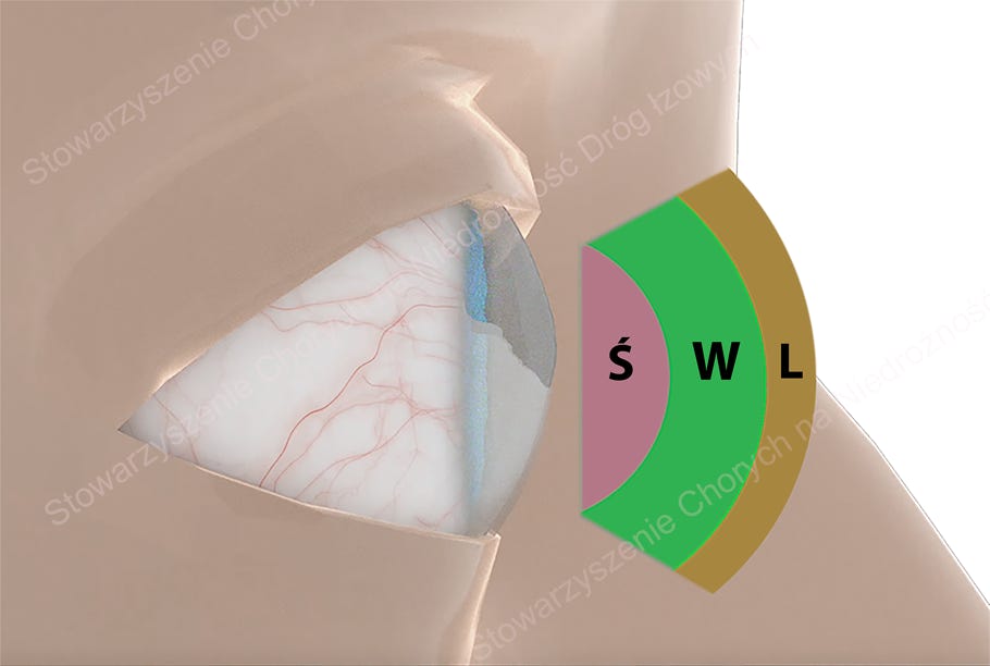
The main lacrimal gland, located in the superolateral part of the orbit, and the accessory glands (Krause, Wolfring, Meibomian, Zeis, and Moll) produce the individual components of the tear film, which serves to protect and hydrate the ocular surface.


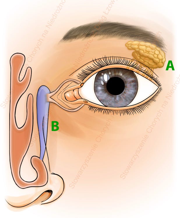
Lacrimal system: A- lacrimal gland; B- lacrimal drainage pathways

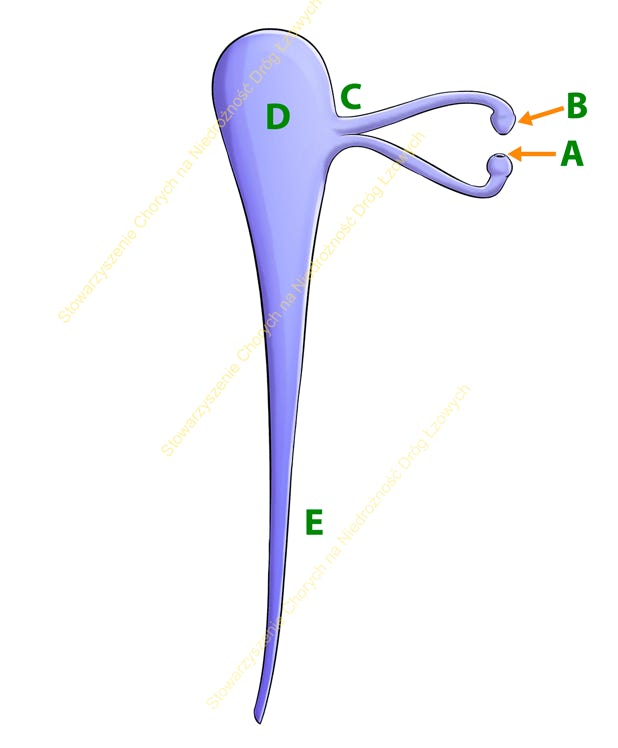
Lacrimal drainage pathways: A- inferior canaliculus; B- superior canaliculus; C- common canaliculus; D- lacrimal sac; E- nasolacrimal duct

Association of Patients with Lacrimal Drainage Obstruction

The Tear Film and Its Layers

The tear film plays a key role in protecting the ocular surface and consists of three layers:

  • Mucin layer – allows adherence of the tear film to the ocular surface.
  • Aqueous layer – provides hydration and transports nutrients.
  • Lipid layer – prevents tear evaporation.

Maintaining a balance between tear production and drainage is essential for ocular comfort and health.


Tear Film: Ś- mucin layer; W- aqueous layer; L- lipid layer

How Do Tears Drain From the Eye?


Tears are spread across the ocular surface by blinking and are then collected by the lacrimal puncta (small openings in the eyelids). From there, they pass through the canaliculi into the lacrimal sac and subsequently drain via the nasolacrimal duct into the nasal cavity.


This system contains natural “valves” that prevent tear reflux. Effective drainage depends on proper eyelid positioning, the functioning of the “lacrimal pump,” and the patency of the drainage pathways.


What Causes Excessive Tearing?


Excessive tearing (epiphora) is most commonly caused by:

  • Tear film abnormalities (e.g., due to chronic blepharitis),
  • Other inflammatory conditions of the ocular surface (e.g., allergic, viral, or bacterial conjunctivitis),
  • Mechanical irritation of the eye (e.g., foreign bodies, eyelashes, eyelid malposition),
  • Impaired tear drainage due to lid malposition (entropion, ectropion), dysfunction of the lacrimal pump (e.g., eyelid laxity), or lacrimal drainage obstruction.

Entropion

Ectropion

Blepharitis and lid laxity

Lacrimal sac abscess

What Is Lacrimal Drainage Obstruction?

Lacrimal drainage obstruction is a condition characterized by partial or complete blockage of the tear drainage pathways from the ocular surface to the nasal cavity.


Under normal conditions, tears produced by the lacrimal glands moisturize the eye and are drained via the lacrimal punctum, canaliculi, lacrimal sac, and nasolacrimal duct. Any obstruction along this pathway leads to tear stasis, resulting in persistent tearing, recurrent conjunctival or lacrimal sac infections, and significant ocular discomfort. In some cases, the inflammation of the lacrimal sac can progress to abscess formation.


Lacrimal drainage obstruction may occur in both children (congenital obstruction) and adults (acquired obstruction) and may be primary (idiopathic) or secondary to inflammation, neoplasms, trauma, or medications.


In most cases, surgical intervention is the treatment of choice to restore proper tear outflow.

Diagnostics of Lacrimal Drainage Obstruction


Diagnostic methods can be classified into basic and advanced evaluations.


Basic diagnostics:

  • Medical history
  • Assessment of the ocular surface and lacrimal outflow system
  • ROPLAS test (pressure over of the lacrimal sac area to check for regurgitation from the puncta)
  • Lacrimal syringing and probing
  • Nasal endoscopy
  • Less commonly used tests: Schirmer test, fluorescein dye disappearance test (FDDT), and Jones tests


Extended diagnostics:

  • Computed tomography - dacryocystography (CT-DCG)
  • Conventional dacryocystography (DCG)
  • Other less common methods: dacryoendoscopy, lacrimal scintigraphy, MRI

CT-DCG: computed tomography scan of the sinus area with imaging of the lacrimal pathways; red arrow points at well filled lacrimal sac and nasolacrimal duct

The selection of diagnostic method is determined by the physician. Extended diagnostics and treatment are typically performed in specialized centers.


Treatment


Initial management focuses on controlling infection and inflammation to prevent complications such as lacrimal sac abscess formation.


In infants with congenital nasolacrimal duct obstruction (NLDO), the first-line treatment is lacrimal sac massage (four times daily, 10 compressions per session). Approximately 20% of infants exhibit signs of congenital NLDO, and over 90% achieve spontaneous resolution or improvement with massage by the age of one year. If conservative management fails, the child should undergo diagnostic - therapeutic probing of the lacrimal system, preferably under general anesthesia with nasal endoscopic guidance.


In adults, surgical intervention is the treatment of choice, depending on the obstruction site:

  • Punctoplasty – for punctal stenosis.
  • Canaliculotomy – incision of the canaliculus, typically for canaliculitis due to dacryoliths (lacrimal stones).
  • Dacryocystorhinostomy (DCR) – for nasolacrimal duct obstruction, involving creation of a bypass between the lacrimal sac and nasal cavity:
    • External DCR (ExDCR): the traditional gold standard; provides good outcomes but may leave a facial scar and requires general anesthesia.
    • Laser-assisted DCR (LDCR): office-based procedure using a laser fiber; less invasive, needs only local anesthesia, but associated with higher recurrence rates.
    • Endoscopic endonasal DCR (EnDCR): the most advanced technique; performed under endoscopic visualization, allowing precise creation of a permanent fistula between the lacrimal sac and nasal cavity without facial scarring.
  • Conjunctivodacryocystorhinostomy with Jones tube – reserved for cases of complete canalicular obstruction; involves implanting a glass tube (Jones tube) to allow tear drainage into the nasal cavity. This method carries a higher complication rate and is used as a last resort.


Video shows technique of endoscopic endonasal dacryocystorhinostomy (EnDCR)

Conclusion:
Regardless of the obstruction type or surgical approach, the decision for operative management should be based on the severity of symptoms—particularly when tearing significantly impairs quality of life or is accompanied by recurrent dacryocystitis.

Association of Patients with Lacrimal Drainage Obstruction

ul. Michala Wolodyjowskiego 21

60-177 Poznan, Poland

KRS (NGO Court Reg. Number): 0000780369

email: lzawienie@icloud.com.wp